he medical artist Pascale Pollier talks in the following interview about how she came to be interested in art and science.
Please tell us about you and your work, and how you came to be a medical illustrator and medical artist.
I was interested in medicine from a very early age. At age 16 I had to choose to either go to art school or to stay on and study science. I was very good at biology and would have loved to work in a laboratory; however, my art teacher recommended I go to art school. This was the first time I had to choose between my two passions, art and science.
I studied fine art and painting at St. Lucas Academy of Art and the Royal Academy of Fine Arts, both in Gent, Belgium, and loved the anatomy lessons and life drawing classes. The subject matter in my paintings was already inspired by medical conditions. I made paintings of hermaphrodites and other medical congenital conditions, including albinism.
On moving to London when I was 19, I saw a job advertised for an artist needed to illustrate a medical encyclopedia. “My dream job!” I thought. When I contacted them, they replied that they could only accept people from the Medical Artists Association, so I contacted the MAA of Great Britain and began my medical art postgraduate training at the University College London. I threw myself into the studies. We drew at the dissection rooms for many hours and studied anatomy from cadavers. It was so wonderful to bring my two passions together at last. I deepened my research into dermatology and endocrinology and made a series of works around my two main themes, albinism and the intersex and third gender.
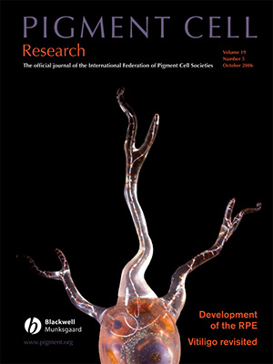
I made a sculpture of a melanocyte in clear resin, which won a prize and it got published on the cover of the Pigment Cell Research Group.
What were some of the most important steps in your career?
In 2001, I moved back to Belgium. It was very difficult to know how to approach the market as a medical artist, as there was no medical art education in Belgium and nobody had even heard about the profession. Professor Gunther Von Hagen was exhibiting in Brussels with the famous Körperwelten. I worked at the exhibition as a medical information officer and had a plastinated lung and a liver at my stand, as well as a slice of a foot and hand. Surrounded by the plastinates and anatomical studies, I made many drawings during break times, and even stayed over all night at the exhibition to draw. I must admit, that was a little eerie.
When the exhibition was finished, I started working as a medical illustrator for a Dutch wholesale pharmaceutical company, specializing in diabetes. They asked me to make an online medical encyclopedia, mapping out the body systems and organs. My dream job! I worked for them for a couple of years, making many paintings and drawings in different media – traditional and digital and a mix of the two.
But even though I was learning a great deal, I was starting to miss the research aspect of my work, the direct contact with scientists, and I felt I hadn’t yet quite found my niche in the medical art field. In 2005, the AEIMS asked me to organize and host their conference in Belgium for 2007. Together with my colleague Ann Van de Velde, a hematologist and medical artist at the University of Antwerp, we organized “Confronting Mortality with Art and Science” at the zoo in Antwerp. For the accompanying exhibition, I made a life-sized wax bust portraying a man, half flayed, examining his own biceps, contemplating his own mortality.
Organizing the conference was a turning point in my career as it made me realize that medical illustration and the pure depiction of functional anatomy was no longer fulfilling my aspirations. The way forward for me as an artist was in collaborating directly with the sciences and to perceive the field of ‘art and science’ in its broadest sense.
By organizing the conference, and the accompanying events like the exhibition and films and book, we had started a network of like-minded people. Collaborating with them to explore the boundaries of art and science was the next, natural step to take.
We also felt there was a real need for artists and student artists to be given the opportunity to study anatomy from the source, to go into laboratories and to engage with scientists. It was in order to help them move forward together into their research fields that we founded BIOMAB Ltd. in 2010. This is an independent, non-profit and non-governmental association offering an interdisciplinary and international program for artists, scholars and scientists. It gave rise, in an inevitable spin-off, to the launch of “Art Researches Science“, which organizes international collaborations of representatives from a wide variety of organizations and courses in New York, London, Dundee, Strasbourg and Antwerp.
At the same time, I had begun working for contemporary artists who needed my expertise and medical knowledge. This led me to become engaged in more sculpture-oriented work. My main materials were now wax, clay, silicone and resin. I was working as a self-employed artist and continued to illustrate books, but was mainly drawn into my studio, making sculptures and organizing and curating exhibitions.
You can find more information on Pascale Pollier and her work under www.artem-medicalis.com.

